Frenectomy and connective tissue graft – Recession treatment case
2014year
7photographs
3sections
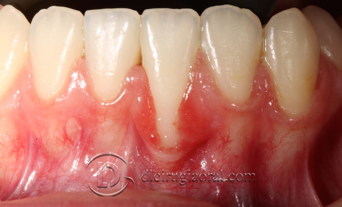
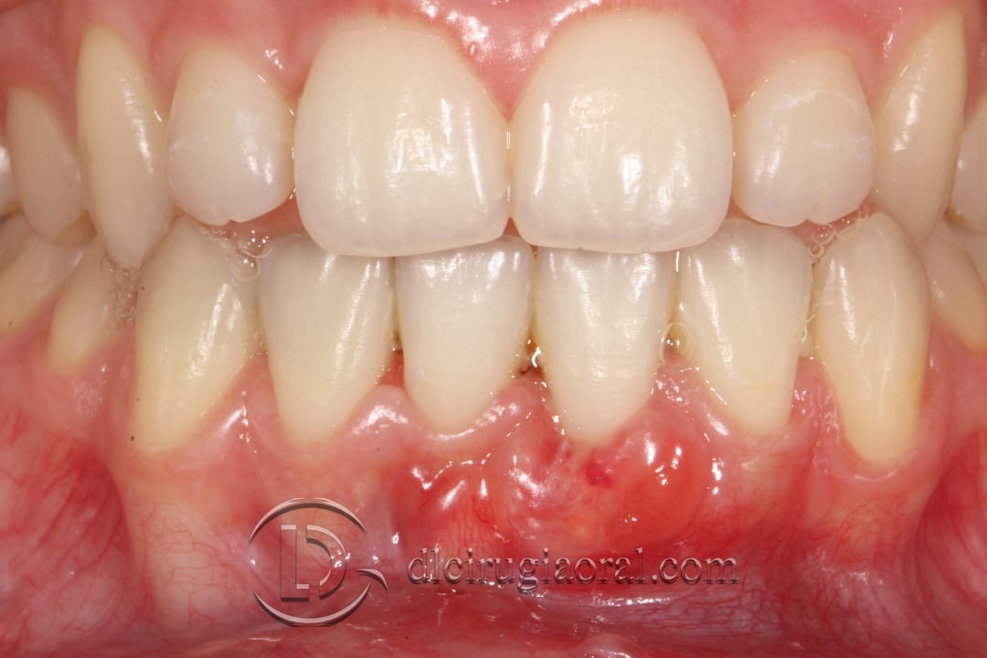
We present the case of a patient referred to us for the management of a gingival recession. She was a non-smoker with no relevant medical history, and presented with a Miller Class II recession affecting a central lower incisor.
Additionally, she exhibited a high frenal attachment and a thin gingival biotype, both of which likely contributed to the recession. These anatomical risk factors are common in patients who have completed orthodontic treatment or present a delicate soft tissue profile.
Combined approach: frenectomy and connective tissue graft
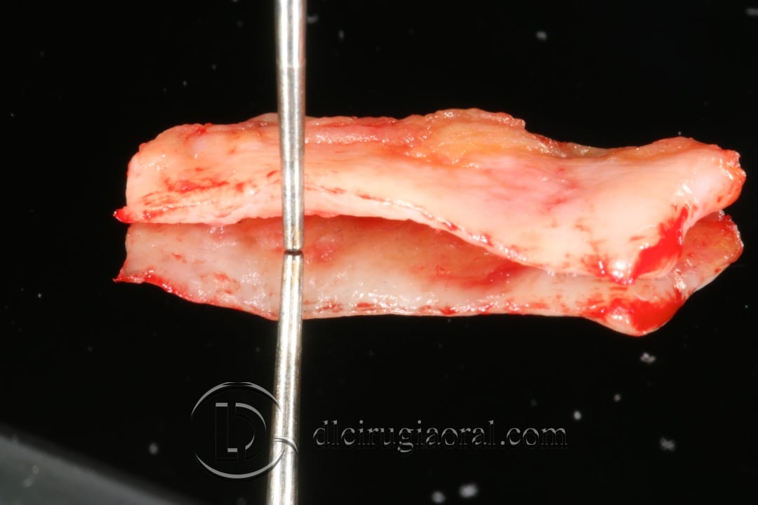
To correct the defect, we chose a combined approach involving frenectomy and connective tissue graft. We first performed a frenectomy to eliminate tension in the area. Immediately afterward, we harvested a connective tissue graft from the palate.
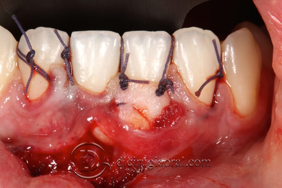
Using a tunnel technique, we inserted the graft into the affected area without raising vertical releasing incisions. This allowed us to preserve the blood supply and minimize trauma. We then secured the graft to the teeth with sutures, following standard soft tissue management protocols.
Healing and clinical outcome
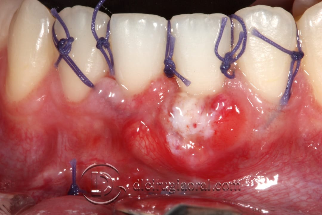
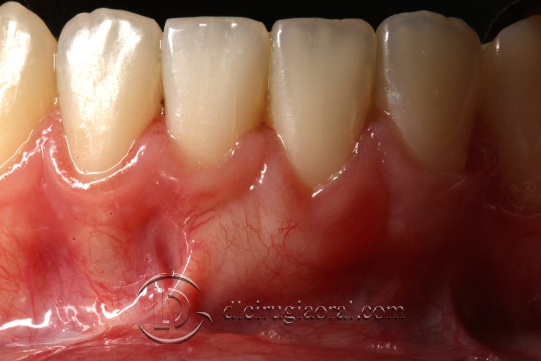
The patient’s healing progressed very well. As seen in the clinical photos, tissue integration and volume improved progressively at one week, 20 days, and 8 weeks post-op.
The outcome showed significant root coverage and a gain in soft tissue thickness, which will help prevent future relapse.
Learn more about recession management
📎 If you want to see more cases involving frenectomy and connective tissue graft, visit our clinical archive for step-by-step documentation.
We hope this clinical case is helpful in your approach to managing thin biotype recessions in the anterior mandible.
Previous situationConnective tissue graftSuturesSituation after 1 weekSituation after 20 daysSituation after 8 weeks