Extraction of Impacted Canine with Immediate Implant and Reconstruction
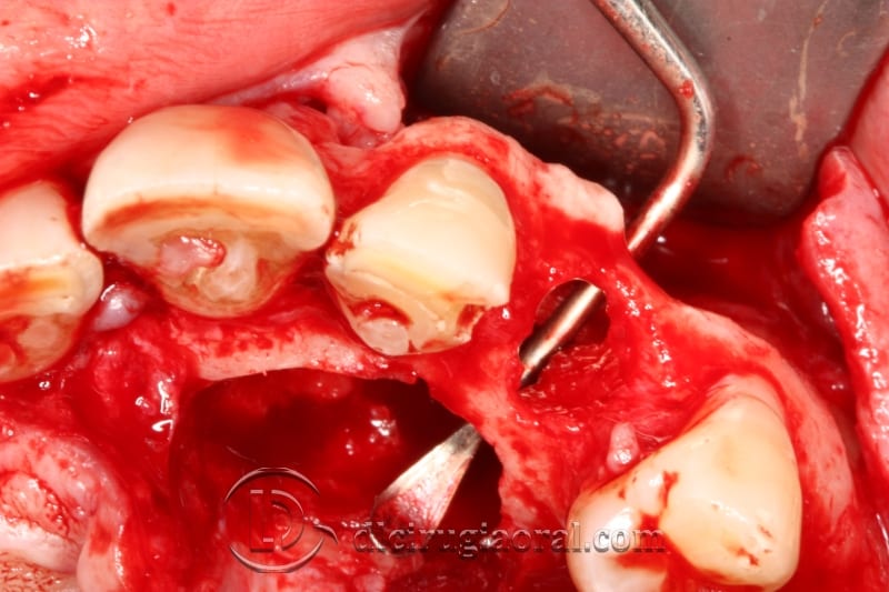
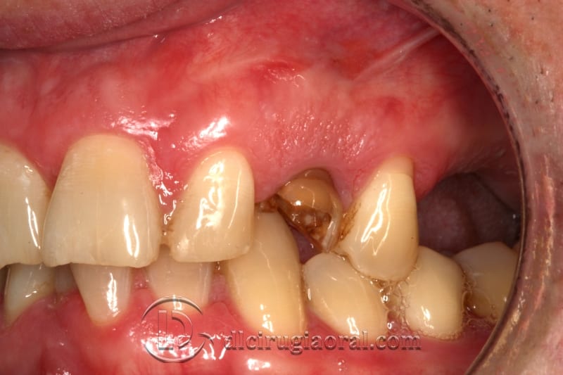
This case details the extraction of impacted canine and immediate rehabilitation through implant placement and autologous bone grafting. The patient, a healthy 35-year-old male, came to our clinic with persistent symptoms in the upper anterior maxilla.
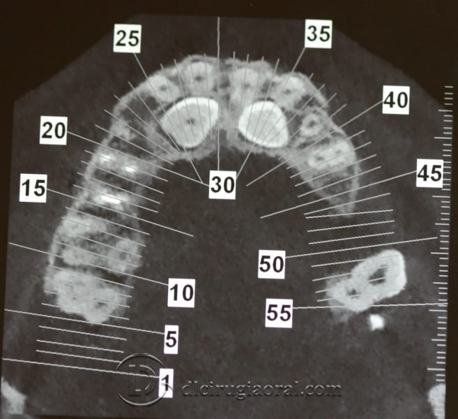
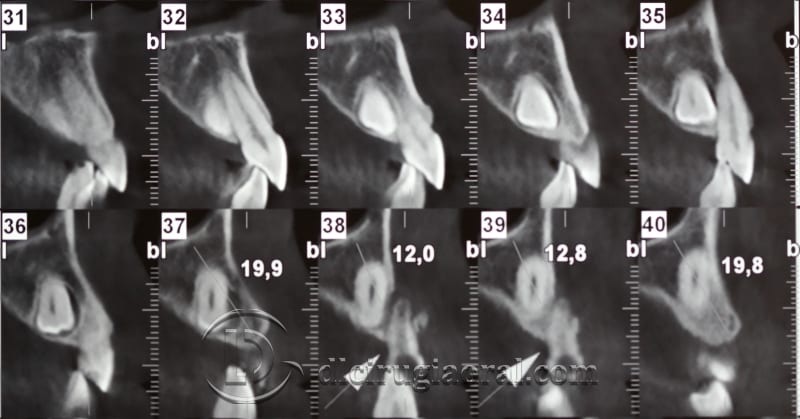
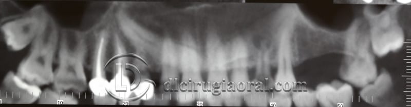
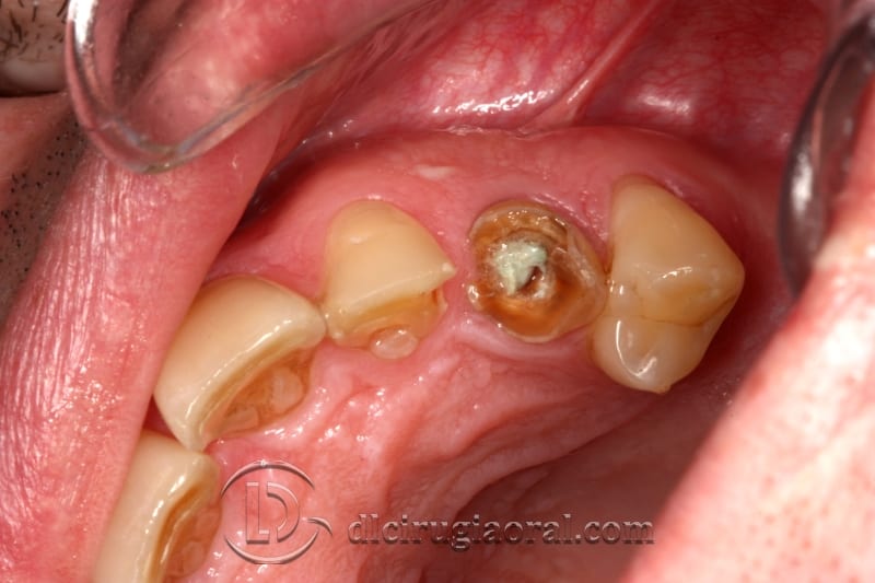
Radiographic evaluation confirmed bilateral impaction of the maxillary canines. Additionally, temporary tooth #63 showed signs of chronic infection and a long-standing fistula.
Choosing a One-Stage Approach
Traditionally, these cases require three surgeries: extraction, bone grafting, and implant placement. That process extends treatment and adds complexity.
In this case, the patient requested fewer interventions. Given his good health, we proposed a one-stage approach: extraction of the impacted canine, implant placement, and bone reconstruction during the same session.
Surgical Strategy and Autologous Grafting
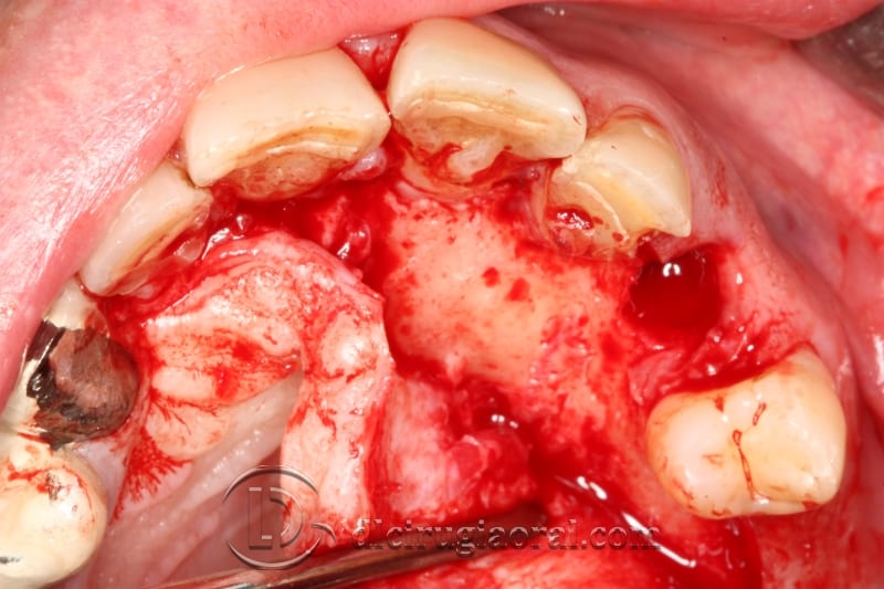
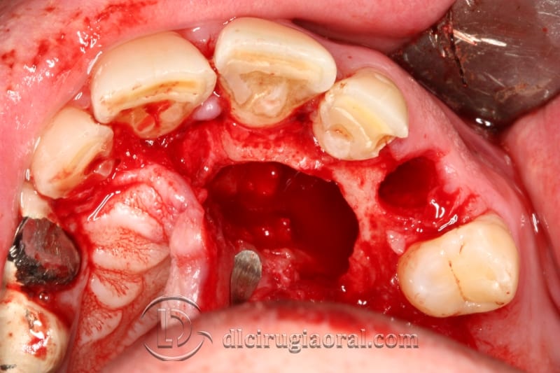
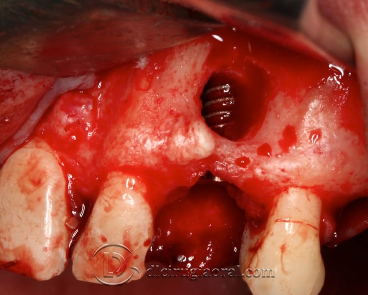
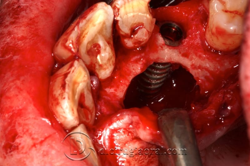
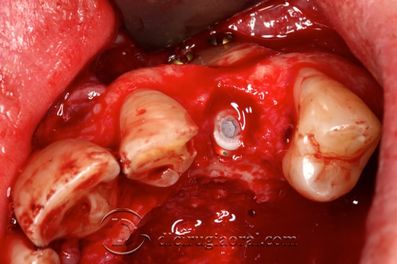
We accessed and removed the impacted canine using a conservative flap. The defect left behind extended buccally and involved palatal and crestal aspects due to the existing infection.
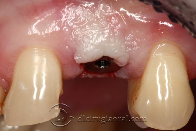
We positioned the implant in site 23, aligned with neighboring teeth and emerging through the cingulum. This orientation supports a prosthetically favorable outcome.
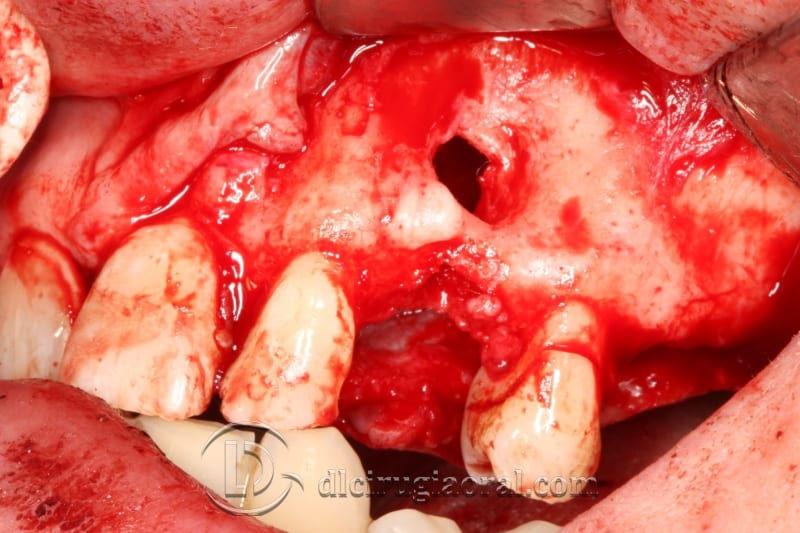
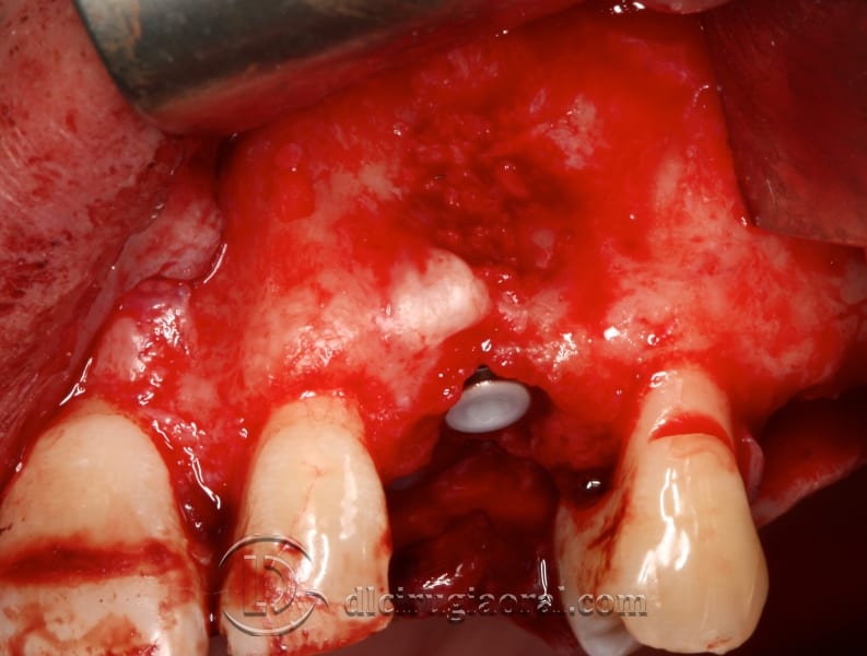
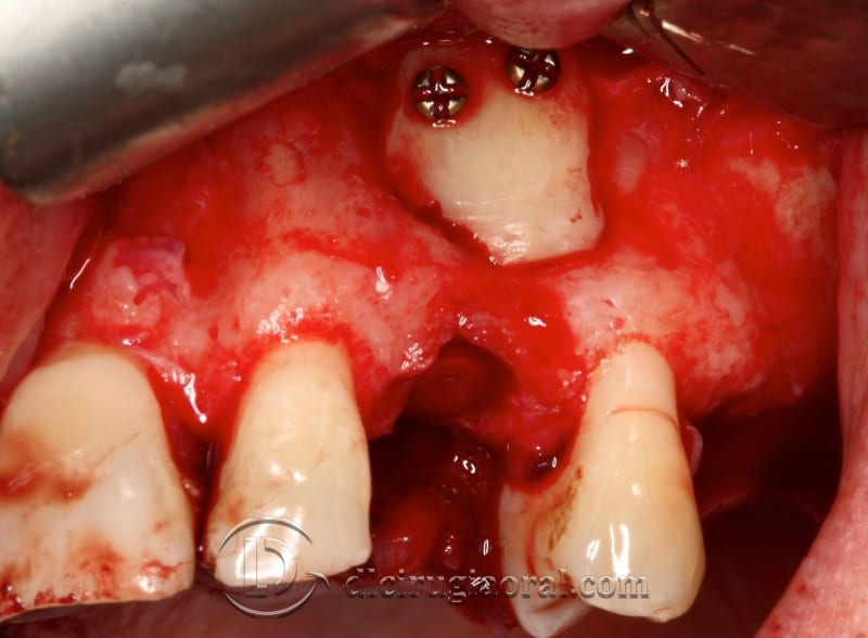
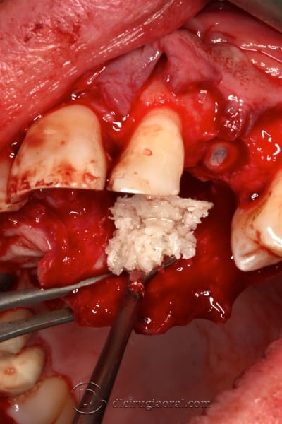
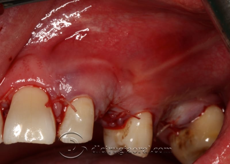
To rebuild the site, we harvested a cortical block from the mandibular ramus. We divided the graft: one half was fixed to the buccal wall with screws, and the other was particulated and packed around the implant. A laminar plate secured the graft, supporting soft tissue closure and healing.
Clinical Outcome and Next Steps
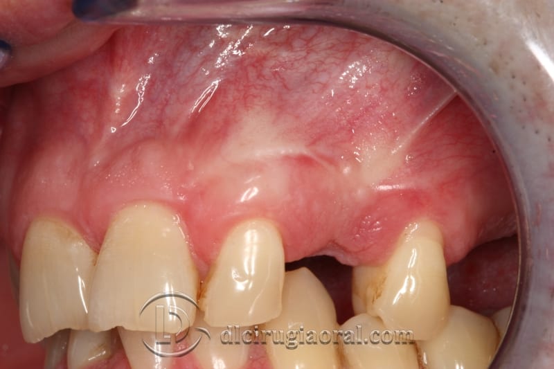
After placing gingival formers, we monitored healing and confirmed tissue stability. Ten days later, we referred the patient to our prosthodontist for the final restoration.
This case shows how planning and proper graft handling can turn a complex extraction into a predictable one-stage treatment.
📎 Learn more: Grafting techniques for fenestrated sites (PubMed)
🔗 See more clinical cases