Atrophy in Central Incisor: Bone Reconstruction and Implant Placement
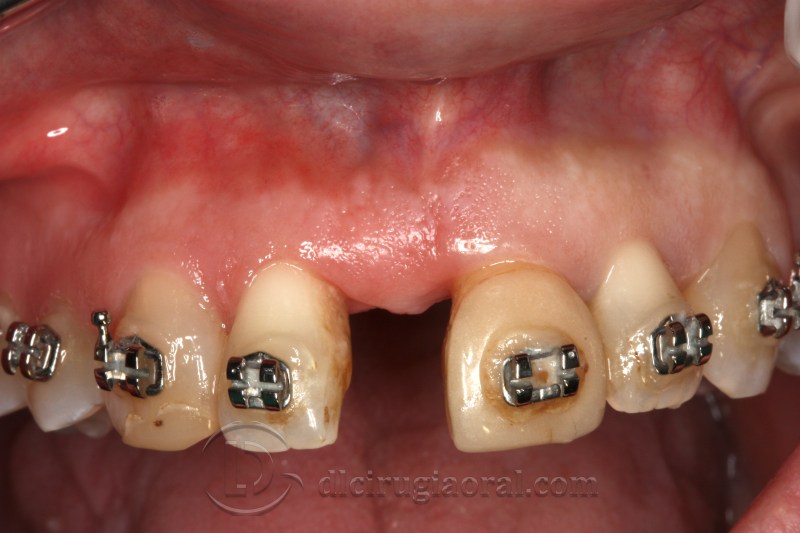
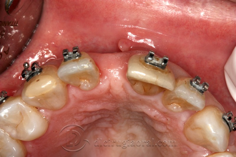
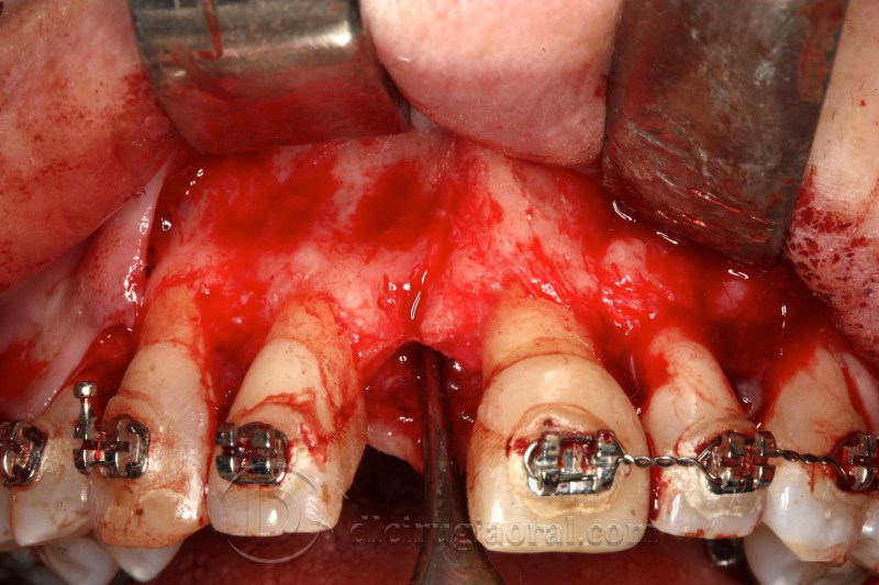
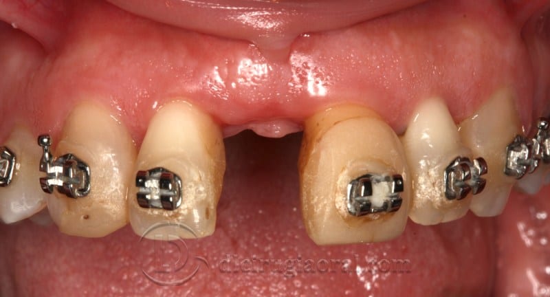
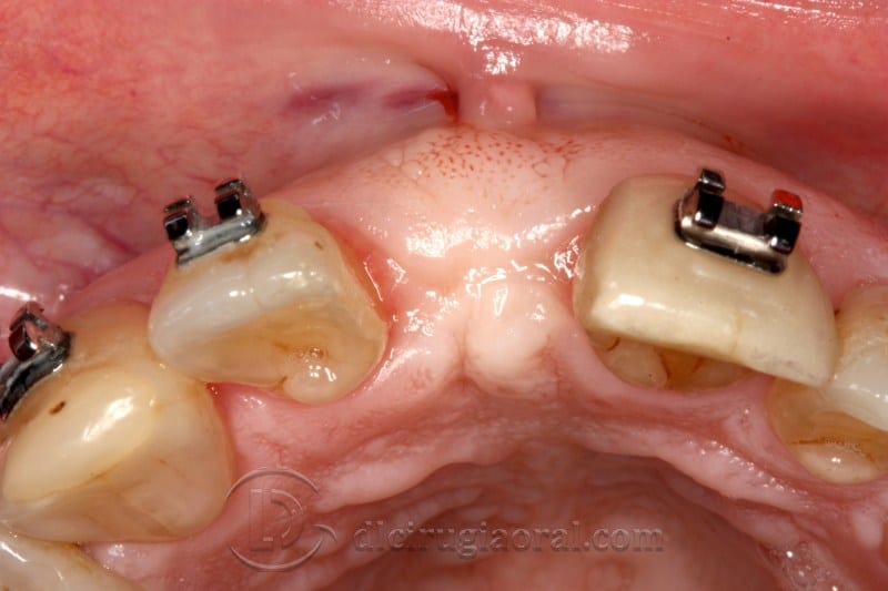
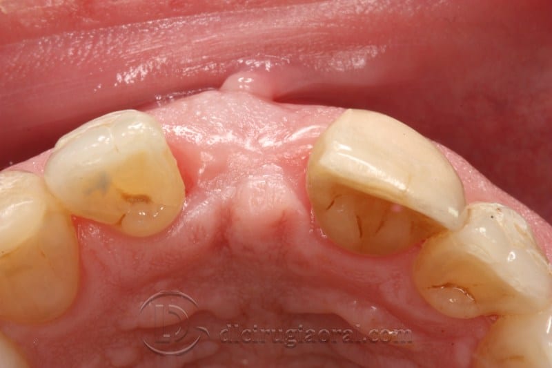
This case focuses on the management of atrophy in central incisor following a childhood trauma. The patient, a 35-year-old non-smoker under Euritox treatment for hypothyroidism, experienced the avulsion of the upper left central incisor during early years. Over time, the alveolar ridge collapsed.
Orthodontic Preparation and Surgical Planning
To correct the malocclusion and reopen the space, the patient underwent orthodontic treatment. She was referred to our clinic at the final stage of orthodontics for implant placement in site 21.
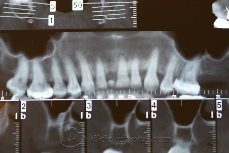
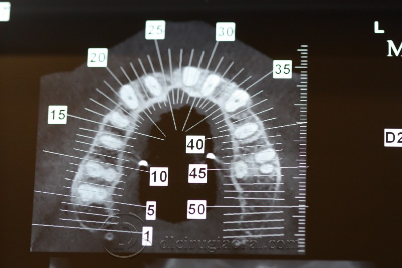
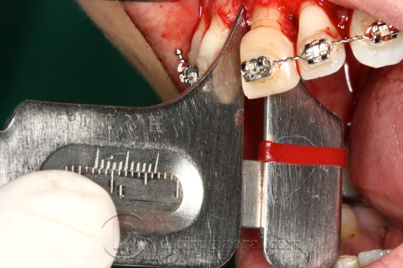
A preoperative CT scan revealed a horizontal bone deficiency. Given the location and the patient’s profile, we planned a staged approach to correct the defect and ensure proper implant integration.
Reconstruction with Mandibular Ramus Graft
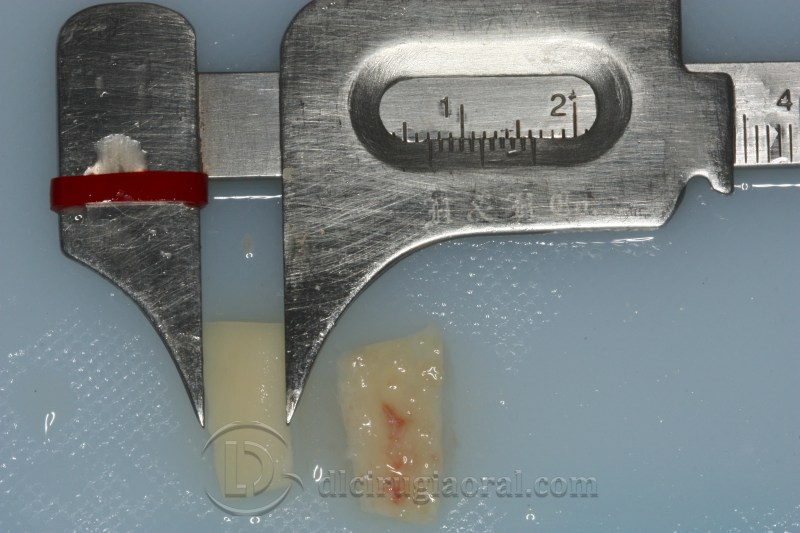
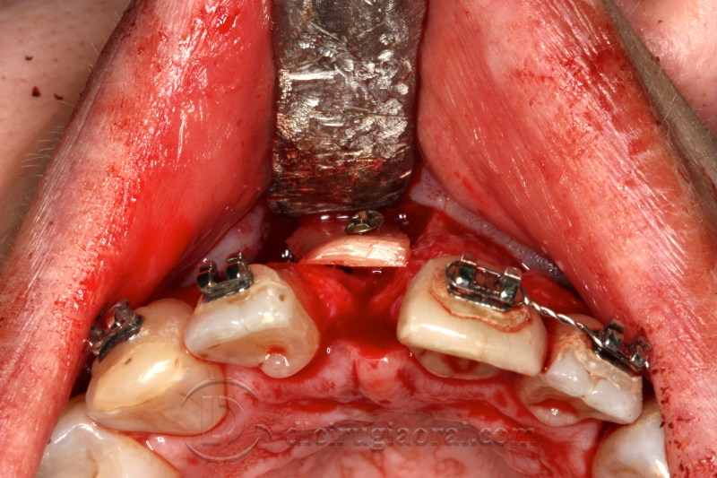
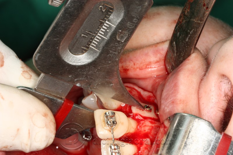
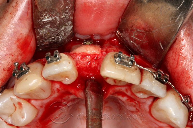
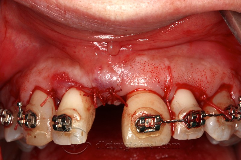
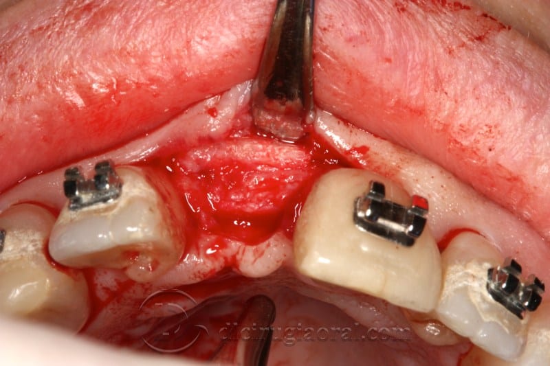
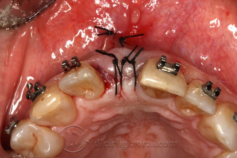
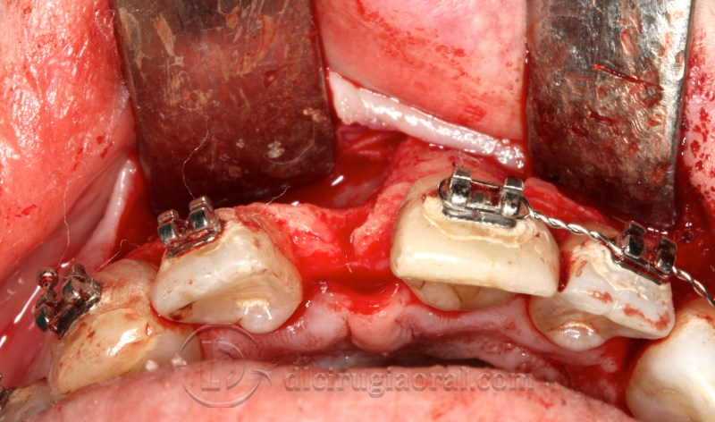
Surgery began with harvesting an autologous graft from the mandibular ramus. We shaped and fixed the bone using osteosynthesis screws to restore the alveolar ridge width. After securing the site, we sutured and allowed a four-month healing period.
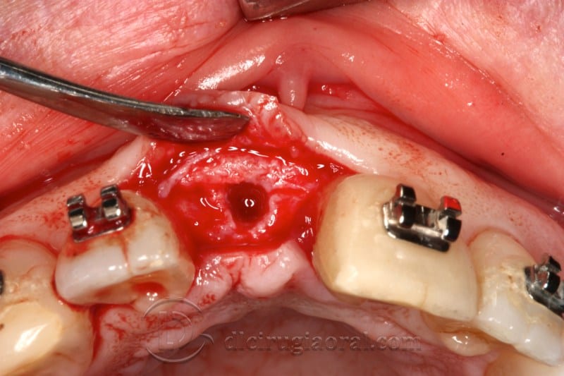
Upon reentry, we observed adequate bone volume and high density during drilling. We then placed the implant in the ideal prosthetic position.
Final Stages and Referral
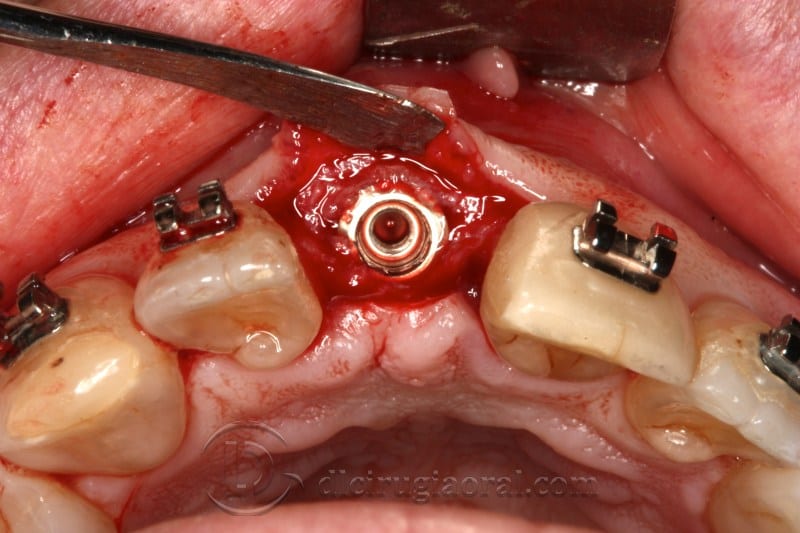
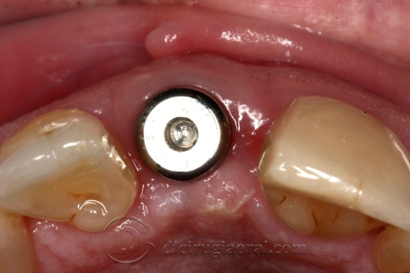
After three months of osseointegration, we placed the gingival former to shape the emergence profile. The patient was then referred to our prosthodontist colleague for the definitive restoration.
📎 PubMed: Bone atrophy and grafting in anterior maxilla
🔗 More anterior grafting cases